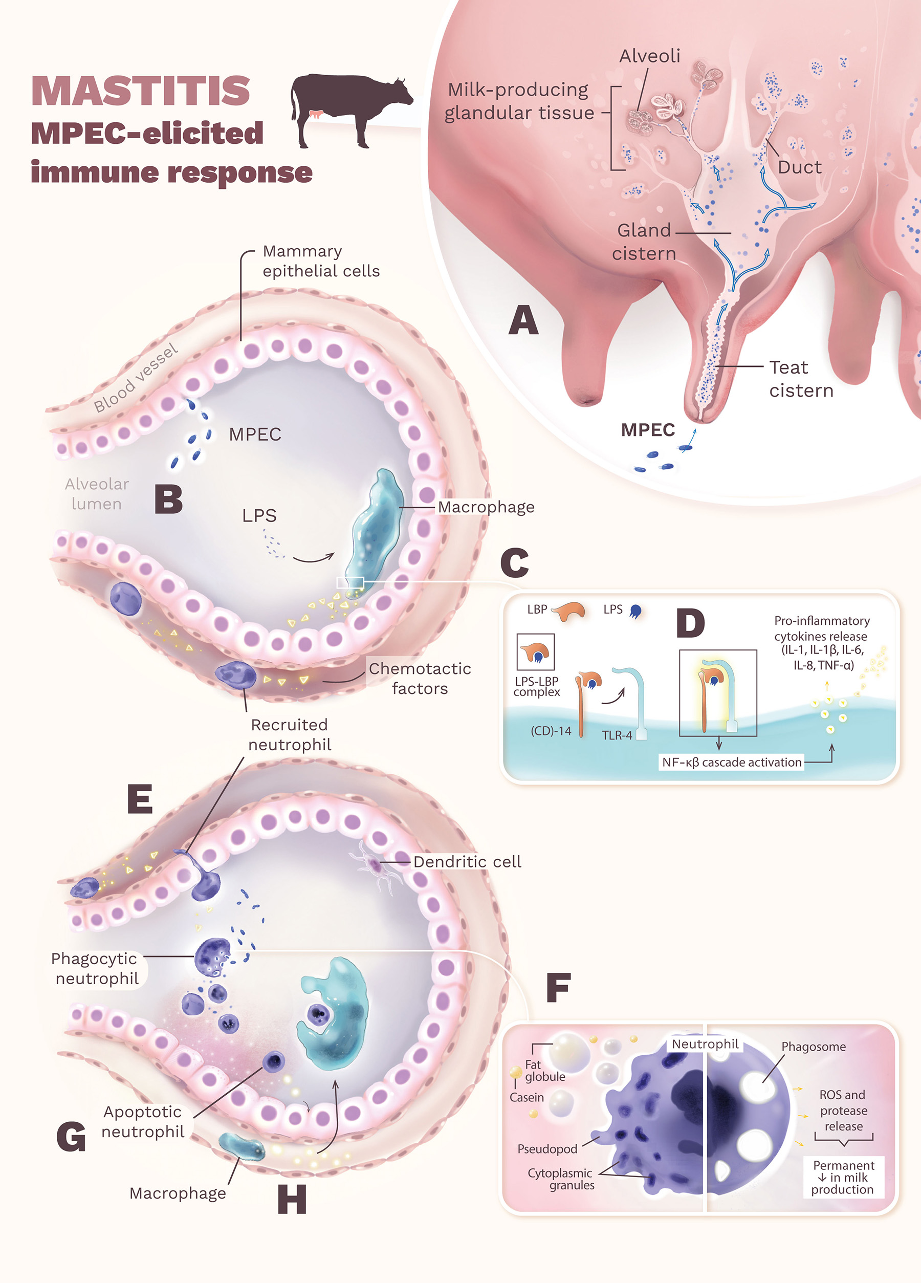
Neutrófilos e macrófagos em ação na resposta à infecção bacteriana que provoca mastite.
Figura produzida para artigo de revisão em periódico científico.
Manuscrito disponível em: doi: 10.3389/fmicb.2022.928346
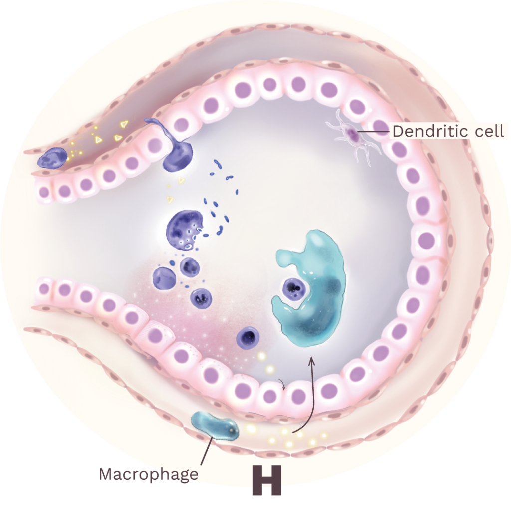
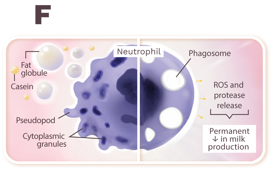
The onset of mastitis occurs when MPEC penetrates through the teat canal, multiplies in the teat and gland cisterns, and spreads throughout the milk-producing glandular tissue (A). The multiplication and lysis of MPEC release LPS from the bacteria’s outer membrane (B). LPS binds to the LBP, and the LPS-LBP complex binds to the CD-14, which leads the LPS to a TLR-4 on the surface of macrophage (C). When LPS binds to its respective TLR, an intracellular signaling cascade is activated (NF-κβ), which induces macrophages and mammary epithelial cells to synthesize pro-inflammatory cytokines and acute-phase proteins (D). Chemotactic factors, especially IL-8, then recruit neutrophils from the bloodstream that function as phagocytes at the site of infection in the alveoli (E). The exposure of neutrophils to fat globules and casein leads to loss of cytoplasmic granules (reduced bactericidal activities) and altered morphology (rounding), eliminating pseudopods needed for phagocytosis (F). During phagocytosis, neutrophils release chemicals that eliminate MPEC but also injure mammary epithelial cells, resulting in a permanent decrease in milk production (F). To minimize mammary tissue damage, neutrophils undergo programmed cell death (apoptosis) and release chemokines that attract macrophages to the site of infection (Paape et al., 2003) (G). In the last step of the inflammatory reaction, macrophages quickly phagocytose apoptotic neutrophils (efferocytosis), minimizing the release of granular contents that damage the tissue (H). Interestingly, whereas in the mammary alveolar, dendritic cells are generally non-responsive to MPEC infection.


